Diagram Of The Muscles In The Forearm - Anatomy Forearm Muscles Anterior Wall Mural - WallMonkeys.com / There are many muscles in the forearm.
Dapatkan link
Facebook
X
Pinterest
Email
Aplikasi Lainnya
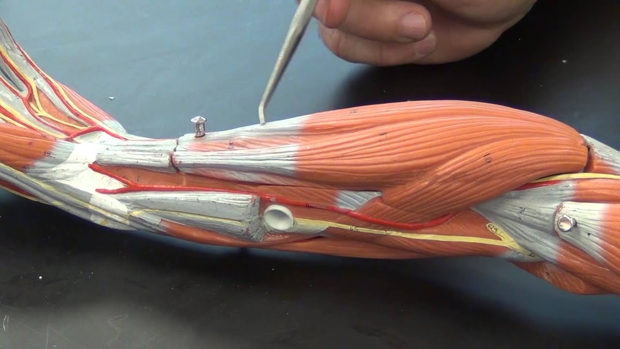
Diagram Of The Muscles In The Forearm - Anatomy Forearm Muscles Anterior Wall Mural - WallMonkeys.com / There are many muscles in the forearm.. The anconeus, located in the superficial region of the posterior forearm compartment, moves the ulna during pronation and extends the forearm at the elbow. Diagram of the forearm extensors superficial extensors consist of seven muscles; Bend your palm toward your forearm. Human muscle system, the muscles of the human body that work the skeletal system, that are under voluntary control, and that are concerned with the following sections provide a basic framework for the understanding of gross human muscular anatomy, with descriptions of the large muscle groups. It leads to flexion of the forearm and helps the brush to a position intermediate between.
The superficial extensors of the forearm are the brachioradialis, extensor carpi radialis longus, anconeus, extensor carpi radialis brevis, extensor carpi ulnaris, extensor digitorum and extensor digiti minimi. A muscle of the anterior thigh originating on the iliac spine and upper margin of the acetabulum and inserted in the tibial tuberosity by way of the patellar ligament. Superficial muscles of the posterior forearm: It leads to flexion of the forearm and helps the brush to a position intermediate between. There are 20 muscles separated into two compartments.
Pictures Of Arm Muscles from healthiack.com The flexor pollicis longus is situated on the radial side of the forearm, lying in the same plane as the preceding. Superficial muscles of the posterior forearm: In the distal forearm, apl and ebp crosses from medial to lateral over ecrl and. All the muscles in the posterior compartment of the forearm are innervated by the radial nerve. Look at the picture of the muscle, find it on your body, and picture how it is contracting as it produces its associated movement or movements. Each muscle roughly follows the course of digits. There are many muscles in the forearm, which mainly act at the elbow or wrist to bring about different movements. Another handy relation to keep in the back of head is:
The flexor digitorum superficialis muscle can be seen underneath these muscles.
I've just switched over to a diagram to show you this muscle. Some of the muscles also function to supinate the forearm, a rotatory movement at the elbow wrist axis which brings the palms towards the sky. Which muscles supinate the forearm? Diagram of the forearm extensors superficial extensors consist of seven muscles; Pronator teres pronates the forearm, turning the hand posteriorly. Here's an example of a petite woman. This muscle connects the humerus to the radius at the styloid process. Bend your palm toward your forearm. The flexor pollicis longus is situated on the radial side of the forearm, lying in the same plane as the preceding. The forearm is a mass of some 20 different muscles. Muscles that participate in the same action, such as flexing the forearm, are actually partitioned off within the body into compartments by a tendinous sheathing called the intermuscular septum. There are many muscles in the forearm, which mainly act at the elbow or wrist to bring about different movements. The superficial extensors of the forearm are the brachioradialis, extensor carpi radialis longus, anconeus, extensor carpi radialis brevis, extensor carpi ulnaris, extensor digitorum and extensor digiti minimi.
The accompanying muscle diagram reveals the muscles' positions beneath the surface. A helpful way to learn anatomy is to move and mimic the actions for the muscles you are learning that week. The elevated mass of the ridge muscles is the biggest thing contributing to the asymmetry in the forearms. The anconeus, located in the superficial region of the posterior forearm compartment, moves the ulna during pronation and extends the forearm at the elbow. The forearm is a mass of some 20 different muscles.
Muscles of the Forearm - YouTube from i.ytimg.com Superficial muscles of the posterior forearm: It leads to flexion of the forearm and helps the brush to a position intermediate between. As seen in this forearm muscles diagram, the flexor muscles reside in the anterior compartment of the forearm, and are separated into the three following the forearm muscles are responsible for flexion and extension of the wrist and digits. Which muscles supinate the forearm? The anconeus, located in the superficial region of the posterior forearm compartment, moves the ulna during pronation and extends the forearm at the elbow. Editor · aug 11, 2017 ·. Your arm muscles allow you to perform hundreds of everyday movements, from making a fist to bending your thumb. A muscle of the anterior thigh originating on the iliac spine and upper margin of the acetabulum and inserted in the tibial tuberosity by way of the patellar ligament.
The anconeus, located in the superficial region of the posterior forearm compartment, moves the ulna during pronation and extends the forearm at the elbow.
Here's an example of a petite woman. Ebraheim's educational animated video describes the anatomy of the supinator muscle. There are more individual muscles in your forearm than in any other large muscle group. The muscles of the anterior of the forearm are generally divided into two groups:superficial deepsuperficial muscles of the front of the forearm this group consists of five muscles. In the anterior compartment, they are split into three categories: The antibrachial or forearm muscles may be divided into a volar and a dorsal group. The 3 muscle groups of the forearm each have their own unique form. Another handy relation to keep in the back of head is: Editor · aug 11, 2017 ·. Look at the picture of the muscle, find it on your body, and picture how it is contracting as it produces its associated movement or movements. The superficial extensors of the forearm are the brachioradialis, extensor carpi radialis longus, anconeus, extensor carpi radialis brevis, extensor carpi ulnaris, extensor digitorum and extensor digiti minimi. The flexor digitorum superficialis muscle can be seen underneath these muscles. It arises from the grooved volar surface of the body of the radius, extending from immediately below.
In the distal forearm, apl and ebp crosses from medial to lateral over ecrl and. A deep layer, intermediate layer and superficial layer. There are many muscles in the forearm. Each muscle roughly follows the course of digits. Learning their anatomy will help you design awesomely dynamic arms.
Diagram Showing Bones Veins And Arteries In A Human Arm ... from media.gettyimages.com The accompanying muscle diagram reveals the muscles' positions beneath the surface. Which muscles supinate the forearm? A muscle lying on the lateral side of the forearm. The forearm is a mass of some 20 different muscles. Superficial muscles of the posterior forearm: There are many muscles in the forearm, which mainly act at the elbow or wrist to bring about different movements. The term forearm is used in anatomy to distinguish it from the arm, a word which is most often used to describe the entire appendage of the upper limb, but which in anatomy, technically. Here's an example of a petite woman.
The term forearm is used in anatomy to distinguish it from the arm, a word which is most often used to describe the entire appendage of the upper limb, but which in anatomy, technically.
This muscle connects the humerus to the radius at the styloid process. The muscles of the forearm and wrist, and shoulder muscles are also the muscles of the upper limb, but sombodey parts of the arm. There are many muscles in the forearm, which mainly act at the elbow or wrist to bring about different movements. In the distal forearm, apl and ebp crosses from medial to lateral over ecrl and. Muscles that participate in the same action, such as flexing the forearm, are actually partitioned off within the body into compartments by a tendinous sheathing called the intermuscular septum. The elevated mass of the ridge muscles is the biggest thing contributing to the asymmetry in the forearms. Here's an example of a petite woman. Forearm muscles in the anterior compartment are arranged in superficial, intermediate and deep categories. Learning their anatomy will help you design awesomely dynamic arms. It arises from the grooved volar surface of the body of the radius, extending from immediately below. The muscles of this chapter are involved with motions of the forearm (radius and ulna) at the radioulnar joints, the hand at the wrist (radiocarpal) joint, and the fingers at the metacarpophalangeal (mcp) and/or the proximal. Each muscle roughly follows the course of digits. Brachioradialis , extensor carpi radialis longus , extensor carpi from the arm muscle diagram above, the muscles of the arm that can be seen easily on the surface include biceps, triceps, brachioradialis, extensor carpi.
Julien Courbet Acteur - Mort De L Acteur Hal Holbrook Les Hommes Du President A 95 Ans - Acteur, producteur, créateur, auteur, scénariste et réalisateur français. . L'animateur vient de perdre vega, une chienne qu'il avait recueillie en refuge. Au printemps 2013, l'animateur quitte mécontent le service public où il officiait sur. Moyenne de tous ses films. Tout ce qu'il faut savoir sur julien courbet. Julien courbet est un acteur français. C'est par hasard que julien courbet a adopté vega il y a des années de cela. 7.2.1965 (55 let) bordeaux, gironde, francie. Il va grandir dans la région et suit des études de commerce à l'iut de bordeaux. Vega, je ne pensais pas que je. Ce mardi 9 février, julien courbet a partagé une triste nouvelle sur son compte twitter. Julien Courbet Info Actualite Potins Mediamass from mediamass.net ...
Camille Kostek Mother : Camille Kostek Height Age Boyfriend Family Biography More : Inside biography 4 camille kostek: . She is the daughter of christina, a gym manager, and alan, a contractor. She first gained recognition for her work with reebok. Camille kostek says modeling agencies told her to lose weight, gronk said don't! Add your childs name on a happy birthday banner, make an easter banner, new year banner, christmas banner, baby shower decoration, congratulations, retirment party banner, mother's day. The model and connecticut native is currently dating nfl star rob gronkowski. Talking about her profession, she started her career. Description of body measurements camille kostek: She is the daughter of christina, a gym manager, and alan, a contractor. Camille kostek and rob gronkowski's romance may just be picture perfect. Camille veronica kostek (born february 19, 1992) is an american model, actress, and host. ...


Komentar
Posting Komentar